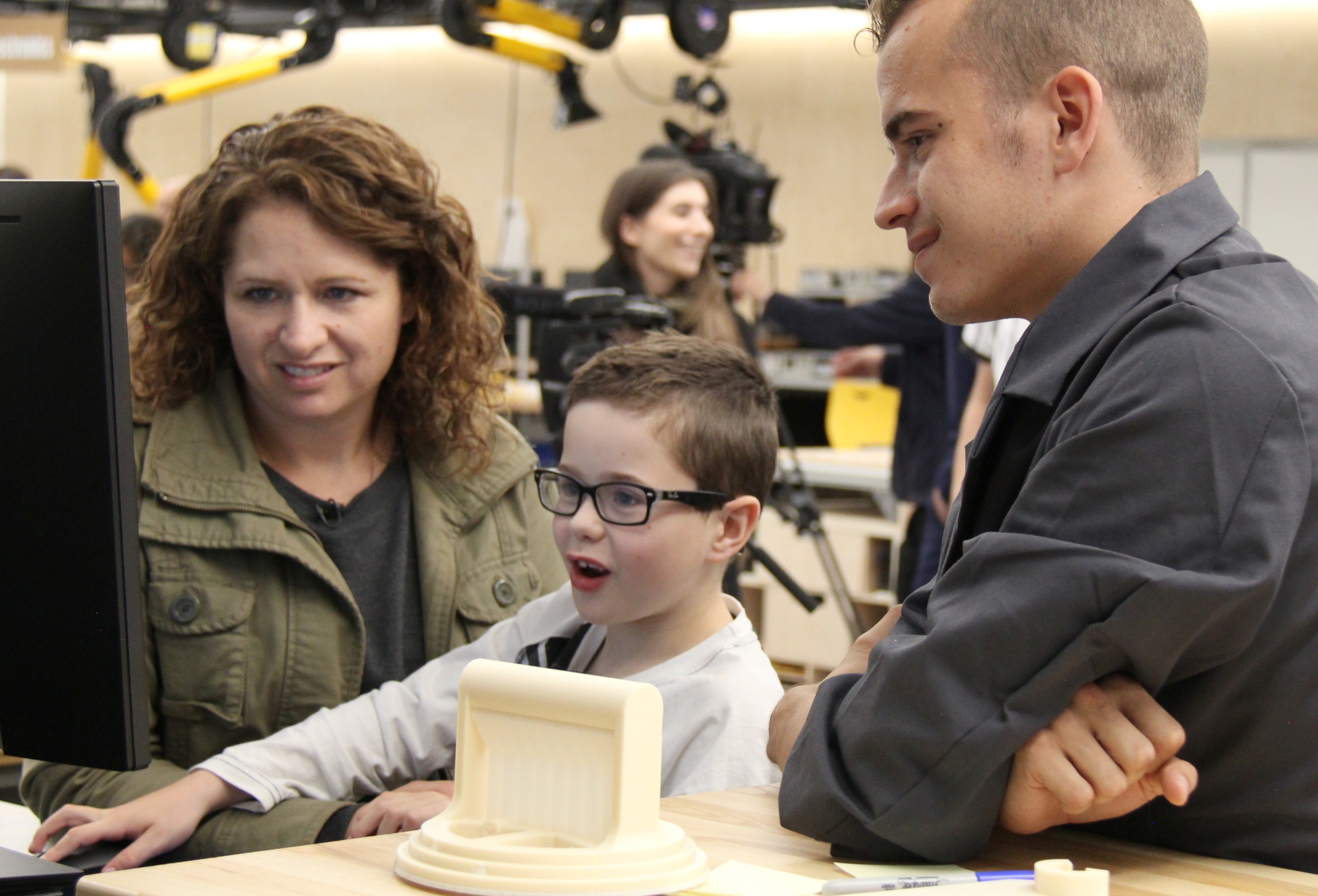
Mason Thomas (centre) with his mother Brandie and engineering student Carter Trautmann look at renderings of Mason's heart on a computer screen. The young heart transplant recipient was presented with 3-D printed replicas of his heart, made through collaborations with doctors, designers, and engineers.
(Edmonton) When Mason Thomas needed a heart transplant at age six, he asked to see his damaged heart so he could understand what was wrong with it, but the old organ was discarded after the surgery.
Two years later, Mason got his wish, thanks to an innovative project by a team of University of Alberta staff and students in medicine, engineering, and industrial design.
Mason received three 3-D models of his heart-one complete with Star Wars TIE Fighter wings-which were designed from CT scan images of Mason's damaged heart and manufactured with a 3-D printer.
"They show why my old heart couldn't work and why I needed a new one," said Mason, now eight. "It's amazing! Thank you so much."
"This is an anatomically accurate representation of Mason's heart, with colours to help him understand how the blood and oxygen were flowing and which parts of his heart were too small and failing," explained project lead Dr. Charles Larson, a U of A clinical lecturer in pediatrics and a pediatric cardiac intensivist at the Stollery Children's Hospital's Pediatric Cardiac Intensive Care Unit, located in the Mazankowski Alberta Heart Institute.
"Mason is a very logical kid who likes to know how things work," said his mother, Brandie Thomas. "The question of why this happened to him bugs him the most, and seeing the heart helps give him some of the answers of what was wrong with him and why he was so sick."
Mason was born with hypoplastic left heart syndrome, a rare and serious congenital defect that was fatal until the 1980s, when surgical fixes were pioneered. Most patients don't need transplants, but Mason's heart failed after his second surgery. He was on oxygen and a feeding tube for the first six years of his life and waited on the transplant list for three and a half years.
Co-operating to create a tactile teaching tool
Larson dreamed up the idea of creating 3-D heart models because it is so difficult for patients, families and medical trainees to visualize congenital heart problems. Doctors will often sketch the heart for patients, but it is hard to capture the complexity in just two dimensions. They also share CT, MRI or ultrasound images, but they are difficult to read for non-experts.
He worked with industrial design students through the Faculty of Medicine and Dentistry's Academic Technologies unit, who took several months to create three-dimensional models by stacking two-dimensional CT scan images on a computer. Then, engineering staff and students at the Elko Engineering Garage, an engineering maker space, stepped up to do the 3-D printing.
Carter Trautmann, an engineering physics student who collaborated with industrial design graduates Cody Wesley and Trina Bloemen, says it was rewarding to apply the high-quality 3-D printing equipment in the Elko Engineering Garage on a medical project.
"It felt petty surreal, almost. To be standing here today to say we made this thing-that is incredible in and of itself, but here's this kid who's getting to see his own heart in a way that would have been unfeasible a few years ago . . . I guess grounding is a good word to describe it," Trautmann said.
"The biggest moment was when I was holding the heart. To be holding it and to see he's missing half, and knowing that this is part of fixing that-that was a little emotional, knowing that this is a thing we can do now."
"None of us in isolation would have been able to create these," Larson said. "It's at the intersection of different fields that breakthroughs happen these days."
Larson noted that donors, such as Servier Canada, supporters of the Servier Virtual Cardiac Centre at the Mazankowski Heart Institute and The Elko family, who helped create the Elko Engineering Garage, and the University of Alberta Hospital Foundation, played essential roles in making this happen.
"We wouldn't have been able to make this happen without engineers and doctors and industrial designers coming together, and likewise, we couldn't have done this without the philanthropy of folks like the Elko family and Servier of Cananda," he said.
"What really surprised and touched me was that the Elko Engineering Garage was open to collaborations," he added. "I'm not an engineer and I came here with an idea from medicine and they said 'That's part of the ethos of this place, is that we want to help other folks and we want to work on collaborations' and I think that is an incredible vision on the part of the Elko family and what has been built here."
Worldwide benefits
The team has created 10 models so far and will make 10 more models of typical congenital heart defects this year. The models will be shared with patients and their families, and will be used as teaching tools to train medical professionals.
Larson said while some groups in the United States are already creating virtual and printed models of damaged hearts for surgical planning, they are usually not based on real patients' scans and are protected by copyright. The U of A team will share the digital models online for free.
"Anyone around the world with a 3-D printer can make these models," Larson said. "A doctor in India with a patient who has the same heart defect could benefit from this work."
Larson said the next step for the project, which is funded by the U of A's Teaching and Learning Enhancement Fund, will be to create models from ultrasound images, which are more complex to read but more commonly available than CT scans. Larson will present the work next month at the World Congress of Intensive Care in Melbourne, Australia.
Brandie Thomas, who works as a paramedic and often speaks to health-care professionals about the cardiac patient experience, believes the models will help young patients cope with their diagnoses.
"People are traumatized when they feel powerless," she said. "If you know exactly what doctors are doing and why they're doing it, it makes you feel that it's being done with you instead of to you and gives you back that sense of control."
As for Mason, he is living as normal a life as possible after his transplant. While he is small for his age and takes immunosuppressants that put him at risk for kidney failure, cancer and infections, he is expected to catch up over time. He loves gymnastics, video games and building things.
"We have a lot of talks before we go to bed about how the heart works and the way the blood flows," said his mother. "He's a tactile learner like me, so it's really meaningful for him to have the model and see his heart for real."
--With files from Richard Cairney