CONNECTIONS Spotlight: Brian Marriott
By Ramona Czakert Franson - 26 August 2022
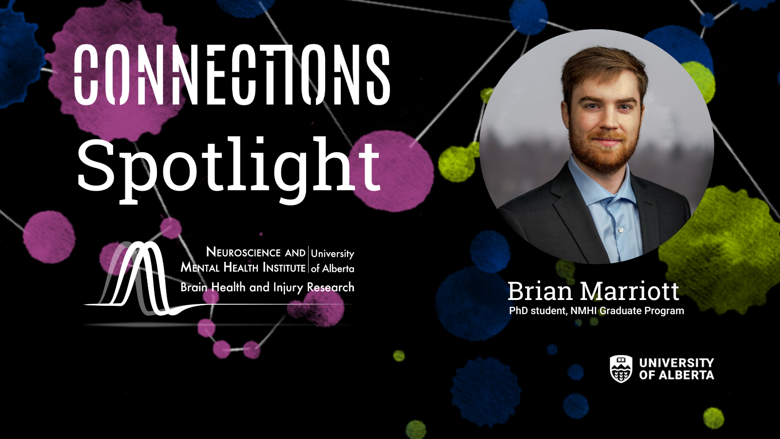
Learn about Brian Marriott, PhD student in the Neuroscience and Mental Health Institute graduate program, and his CONNECTIONS images. In his research, Brian uses microscopy to study a unique part of the brain called the claustrum. By bringing his passions for microscopy and photography together, he has created three visually stunning images of the brain for CONNECTIONS, NMHI’s gallery project that brings neuroscience and art together. We asked Brian to elaborate on these images.
Tell us about your CONNECTIONS images, Nexus, Aurora and Plastic Brain.
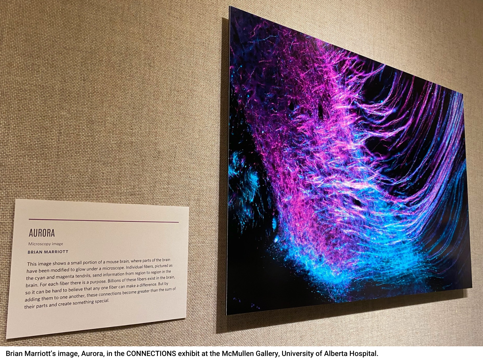
In Aurora, a mouse was injected with two special viruses in one region of the brain, revealing all the different parts of the brain that are connected. It’s like looking at a city on a road map and highlighting all the direct roads that lead to it. In the image, you are directly seeing the pathways the brain uses to connect and send information from one region to another.
In Nexus, the different coloured circles are all neurons in a region of the brain called the claustrum. The reason they’re different colours is because this 2D image is a representation of a 3D volume of brain tissue. The neurons in red are at the top of this volume, and the colours progress through to blue which is at the bottom. This colour coding is one way to provide spatial information in an otherwise 2D image, which is important when the brain itself is a 3D object.
Plastic Brain is a self-portrait where I’m holding a 3D printed brain cell called an astrocyte. Astrocytes are quite different to neurons, but they’re equally important in the brain as they support brain function and health. The colours illuminating this subject were chosen to mirror the colours of light we often use or see when using microscopes to obtain images.
What is your work/research/studies about?
My research is focused on the claustrum. The claustrum is a small, curved sheet of neurons under the outside layer of the brain that has been difficult to study using conventional techniques. This region of the brain has an incredible number of connections going to and from it, which have been well studied; however, the actual function of the claustrum isn’t fully understood. My research looks at the nuances of how individual claustrum neurons are organized and connect to other areas of the brain, and at how the claustrum is active across waking and sleep. Other research has shown that generally speaking, the claustrum is highly active during sleep, but my goal is to dive deeper and show what individual claustrum cells are doing during sleep.
Why the University of Alberta and neuroscience?
During my undergrad at Mount Royal University in Calgary I was fortunate to have the opportunity to do undergraduate research with an amazing supervisor Dr. Adrienne Benediktsson, who infected me with a love of neuroscience and microscopes! She showed me Golgi stained brain slices under the microscope, and seeing the beautiful complexity of the brain up close made me want to pursue neuroscience further.
After I finished my undergraduate degree, I began looking for a master’s degree supervisor that aligned both with my microscopy experience and neuroscience focus. In my search, I found U of A’s Dr. Jesse Jackson (my current supervisor). I was very interested in the cutting-edge techniques he was proposing for his brand-new lab, which have turned out to be essential to the work I’ve been doing. And crucially, he agreed to take me on as a student!
Is the brain beautiful?
Absolutely! Interestingly, the neuroanatomist Ramon Y Cajal, in the 1880-1900’s had to painstakingly paint and draw what he saw under the microscope. His works are both scientific and artistic in the representation of the brain. The tools have changed how we view the brain, but I think, just as it was over 100 years ago, the brain is truly captivating under the microscope, and I’m grateful for the opportunity to bring it out of the lab.
More about CONNECTIONS
Launched by the Neuroscience and Mental Health Institute (NMHI), in collaboration with the Faculty of Arts, CONNECTIONS is a project showcasing the relationship between art and neuroscience, portraying the many aspects of neuroscience, brain diseases and mental health in a creative and compelling way. Artists, scientists and persons with relevant lived experience were asked to submit artwork that related to this theme.
The NMHI is a multi-faculty, interdisciplinary teaching and research institute located at the University of Alberta. Our members come from medicine, pharmacy, science, rehabilitation medicine, kinesiology, psychology, nursing, business, engineering, computing science, arts, and more.
Aside from showcasing the relationship between art and neuroscience, the CONNECTIONS project will help to support research at the NMHI.
Follow NMHI’s Facebook and Twitter for posts that spotlight works from scientists and artists from many different departments and faculties at the U of A. And an on-demand book to purchase is coming soon!