Neuroscience: A Journey Through the Brain
|
Neuroscience: A Journey Through the Brain |
| Main Page Organization Development Neuron Systems About the site (Glossary) References |
The Major Systems: Sensory Systems - Somatosensory System
|
Sensory System sub categories: Chemical Visual Auditory Somatic |
To understand how humans perceive somatic sensations, we must explore the following:
Types of Somatosensory Receptors (back to top)
| Category | Receptor | Function |
| Mechanoreceptors | Pacinian corpuscle | Fast adapting with large receptive fields for signifying the beginning and end of gross somatosensations. |
| Meissner's corpuscle | Fast adapting with small receptive fields to signify the beginning and end of fine somatosensations. | |
| Merkel's disk | Slow adapting with small receptive fields to signify sustained and fine somatosensations. | |
| Ruffini's ending | Slow adapting with large receptive fields to signify sustained and gross somatosensations. | |
| Nociceptors | Mechanical nociceptor | Respond to strong pressure from sharp objects. |
| Thermal nociceptor | Respond to burning heat or extreme cold. | |
| Chemically sensitive, mechanically insensitive | Respond to extremes in pH and to irritants. | |
| Polymodal nociceptors | Respond to combinations of mechanical, thermal, and chemical stimuli. | |
| Thermoreceptors | Warm receptors | Respond to temperatures between 30 and 45 °C. |
| Cold receptors | Respond to temperatures between 10 and 35 °C. | |
| Proprioceptors | Muscle spindles | Measure the length and rate of stretch of muscles. |
| Golgi tendon organs | Gauge the force generated by a muscle by measuring the tension in the tendon. |
Somatosensory Pathways (back to top)
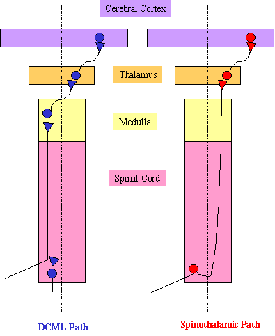 |
Somatic sensory signals can take two major routes to the CNS. The dorsal column-medial lemniscal pathway carries information about touch and vibration from the skin, as well as proprioceptive information from the limbs. The spinothalamic pathway carries information about pain and temperature. The projections of the two pathways are depicted to the left. The two pathways segregate at the margin of the spinal cord. The DCML path axons synapse on the ventral posterior nucleus (VPN) of the thalamus. The VPN then projects to the primary somatosensory cortex or S1. The spinothalamic neurons also synapse on VPN neurons, but in a different region. They also synapse on the small intralaminar nuclei of the thalamus. The big difference between the two is the level of decussation - the DCML path decussates at the level of the medulla, while the spinothalamic path decussates much earlier in the spinal cord. The DCML path involves large diameter fibers, and the spinothalamic path involves small diameter fibers. Large diameter fibers enter more medially, and small diameter fibers enter more laterally, through the Zone of Lissauer. Large diameter fibers send one branch to the dorsal horn and one branch to the dorsal column on the same side. Small diameter fibers send branches to the dorsal horn, which then sends an axon across the cord, and into the spinothalamic tract. |
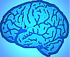
Somatosensory Cortex (back to top)
Most of the cortex concerned with somatosensation is located in the parietal lobe. Primary somatosensory cortex (S1) occupies an exposed cortical strip called the post central gyrus, which lies just posterior to the central sulcus. S1 is divided into four distinct regions: Brodmann's areas 3a, 3b, 1 and 2, with the order starting from the central sulcus and moving backwards. Area 3b is concerned with texture size and shape of objects, and projects texture information to area 1, as well as size and shape information to area 2. Secondary somatosensory cortex (S2) can be revealed by pulling back the temporal lobe and looking at the lower part of the posterior lobe. The posterior parietal cortex consists of Brodmann's areas 5 and 7, and sits just posterior to S1.
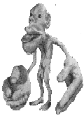
The primary somatosensory cortex contains a somatotopic map of the body's surface:
 |
Like the auditory tonotopic map, the somatotopic map means that adjacent parts of the body are mapped onto adjacent parts of the primary somatosensory cortex. We can see from the illustration that an improprotinately large amount of cortex is devoted to the fingers and to parts of the face like the mouth. The following cartoon illustrates what a human would look like if the amount of cortex were proportionate to the size of the organ or limb:
|
Created and Maintained by: Melissa
Davies
Last Updated: April 09, 2002 09:02 PM