Ectosomes: Tiny Bubbles, Big Impact
Dr. Julie Jacquemyn - 29 May 2026
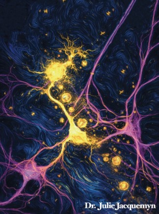
Image by Dr. Julie Jacquemyn
Parkinson’s disease is a progressive neurodegenerative movement disorder that affects 10 million people worldwide, including over 100,000 Canadians. Parkinson’s disease is highly complex and often described as a multifaceted disorder because no two patients experience it in exactly the same way. This complexity stems from the degeneration of dopamine-producing neurons inside the brain, leading to a broad spectrum of motor (tremor, imbalance, muscle cramps) and non-motor symptoms (depression, anxiety, sleep disturbances) that vary considerably in both type and progression. This loss can result from genetic mutations, environmental factors (like pesticide exposure), and/or aging.
A key feature of Parkinson’s disease is the accumulation of misfolded alpha-synuclein. It’s a protein that normally plays a very positive role in the brain, controlling the release of chemical messengers that cells use to communicate. In Parkinson’s disease, however, alpha-synuclein misfolds and clumps together creating large aggregates. These aggregates are toxic to neurons and ultimately will lead to cell death. Mounting evidence also shows that alpha-synuclein can spread from one brain cell to another, contributing to disease progression. Therefore understanding the core mechanism(s) of spread is a central challenge in the Parkinson’s disease field that needs to be resolved.
In addition to alpha-synuclein misfolding, fat alterations also play a major role in Parkinson’s disease. Mutations in Parkinson’s disease risk genes cause certain fat molecules to build up and disrupt normal cell function. This buildup is linked to more alpha-synuclein clumping. But how these fats influence the spread of alpha-synuclein between cells is still not fully understood.
One idea is that these fats trigger the release of tiny bubbles called extracellular vesicles, which can transport alpha-synuclein between cells. However, cells produce many different types of these vesicles and it was unclear which type is responsible for transmitting alpha-synuclein.
Dr. Maria Ioannou and Dr. Julie Jacquemyn are helping us to uncover this mystery. In their most recent publication, they investigated how alterations in brain fats contribute to the formation of extracellular vesicles. They explored this using dopaminergic neurons derived from cells obtained from different Parkinson’s disease patients. They discovered that changes in fats called sphingolipids, cause Parkinson’s disease cells to release a subtype of vesicle called ectosomes. These ectosomes bud from the cell surface, carry harmful α-synuclein and can pass it to nearby cells. And that restoring sphingolipid levels prevented ectosome formation and spread of disease pathology from one neuron to the next.
Overall, this study shows that changes in cell fats linked to common Parkinson’s disease genes may help drive the spread of harmful proteins between brain cells. This makes ectosomes a potential target for future treatments aimed at slowing disease progression. However, many questions are still unanswered. Dr. Maria Ioannou and Dr. Julie Jacquemyn continue to investigate how ectosomes form and how proteins like alpha-synuclein are selected and packaged into them. It also remains unclear how this pathway compares to other possible routes of protein spread, as multiple mechanisms may act together depending on brain region and cellular conditions. Together, this ongoing research will be key to better understanding how Parkinson’s disease spreads and to developing new strategies to stop it.
So, tiny bubbles do have a big impact in Parkinson’s disease!