CONNECTIONS Spotlight: Chinmayee Das
By Ramona Czakert Franson - 16 September 2022
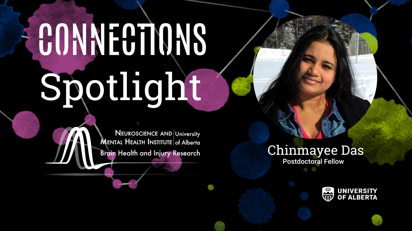
Learn about Chinmayee Das, a postdoctoral fellow working with Neuroscience and Mental Health Institute member Simonetta Sipione, and her CONNECTIONS images. Using microscopy, Chinmayee has created two visually stunning images for CONNECTIONS, NMHI’s gallery project that brings neuroscience and art together. We asked Chinmayee to tell us more.
Questions:
Tell us about your CONNECTIONS images An Angle of Observation and Eustress or Distress?
My work is a collection of immunofluorescent images indicative of neural network and calcium-sensing receptors around the eye of a two-day-old zebrafish larvae exposed to acute stress. It is a step in understanding the visual perception of danger and stress. Calcium rushes into the cells when the eye perceives a threat, and it triggers a “fight or flight” response, such as rapid muscle movement to be able to run away from the danger, or to be aware of the surroundings and hide. Live larvae imaging is an efficient tool to look at the fastest possible responses happening during stress. My PhD project on acute stress is my inspiration for the images using microscopy as a crucial tool to visualize rapid changes in zebrafish larvae. Imaging has given me perspective and helped me understand the concept better.
What is your work/research/studies about?
My current research investigates the role of lipid droplets in neuronal proteostasis. Proteostasis also known as protein quality control is essential for the proper functioning of every cell, in particular, brain cells. Failure of this control network leads to the accumulation of protein waste inside the cells, triggering toxicity followed by cell death. My goal is to understand the actions taken by brain cells during such failures to protect and preserve cell functions and health. My work is focused on understanding the potential role of cellular lipid droplets (LDs) as the first line of defence against the accumulation of toxic protein waste. Lipid droplets are made of fatty substances and might present a “sticky” surface that could trap protein waste. Using biochemical and imaging approaches, I am trying to elucidate how LDs may help in reducing the load of toxic protein waste and how they might help brain cells get rid of this waste.
Why the University of Alberta and neuroscience?
I am curious about the brain and its various facets and I am very grateful for the opportunity to work at the U of A. The U of A has excellent core facilities and I have learned new skills working here. I am a postdoctoral fellow working in collaboration with Richard Lehner and Simonetta Sipione.
Is the brain beautiful?
The brain is the most complicated yet beautiful organ. It is like chambers within chambers and each leaflet of the chamber leads to a different finding! How fascinating! I am intrigued by different pathways that coordinate multiple functions at the same time in such an efficient way. I strongly believe the brain is the most beautiful organ.