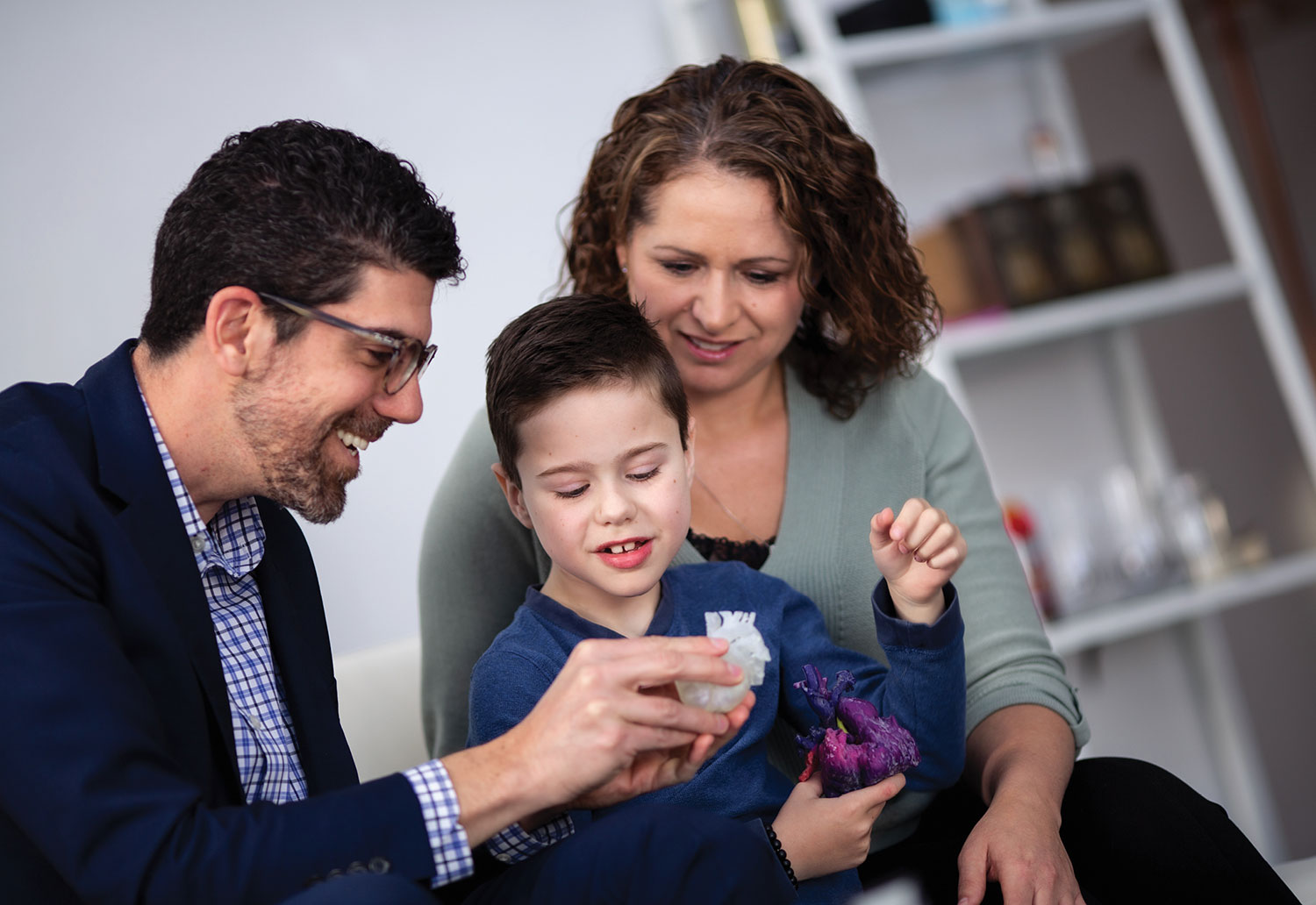
"They show why my old heart couldn't work and why I needed a new one. It's amazing!" - Mason Thomas.
Photo: Laughing Dog Photography
When Mason Thomas needed a heart transplant at age six, he asked to see his damaged heart so he could understand what was wrong with it, but the old organ was discarded after the surgery.
Two years later in the fall of 2019, Mason got his wish, thanks to an innovative project by a team of University of Alberta staff and students in medicine, engineering and industrial design.
Mason received three 3D models of his heart-one complete with Star Wars TIE Fighter wings-which were designed from CT scan images of Mason's damaged heart and manufactured with a 3D printer.
"They show why my old heart couldn't work and why I needed a new one," Mason said when he received the models. "It's amazing! Thank you so much."
"This is an anatomically accurate representation of Mason's heart, with colours to help him understand how the blood and oxygen were flowing and which parts of his heart were too small and failing," explains project lead Charles Larson, MD. Larson is a member of the Women and Children's Health Research Institute, U of A clinical lecturer in pediatrics and pediatric cardiac intensivist at the Stollery Children's Hospital's Pediatric Cardiac Intensive Care Unit.
"Mason is a very logical kid who likes to know how things work," says his mother, Brandie Thomas. "The question of why this happened to him bugs him the most, and seeing the heart helps give him some of the answers of what was wrong with him and why he was so sick."
Mason was born with hypoplastic left heart syndrome, a rare and serious congenital defect that was fatal until the 1980s, when surgical fixes were pioneered. Most patients don't need transplants, but Mason's heart failed after his second surgery. He was on oxygen and a feeding tube for the first six years of his life and waited on the transplant list for three and a half years.
Co-operating to create a tactile teaching tool
Larson dreamed up the idea of creating 3D heart models because it is so difficult for patients, families and medical trainees to visualize congenital heart problems. Doctors will often sketch the heart for patients, but it is hard to capture the complexity in just two dimensions. They also share CT, MRI or ultrasound images, but they are difficult to read for non-experts.
"Your heart is inside your chest walls and you never see it," Larson says. "The plumbing is very complex."
Larson worked with industrial design students through the Faculty of Medicine & Dentistry's Academic Technologies unit, who took several months to create three-dimensional models by stacking two-dimensional CT scan images on a computer. Then staff and students at the Elko Engineering Garage, an engineering faculty workshop, stepped up to do the 3D printing.
"None of us in isolation would have been able to create these," Larson says. "It's at the intersection of different fields that breakthroughs happen these days."

Following a heart transplant at age six, Mason Thomas was given 3D models of his heart to help him understand what made the procedure necessary.
Photo: Laughing Dog Photography
Worldwide benefits
The team has created 40 models so far and will make more models of typical congenital heart defects this year. The models are shared with patients and their families, and are used as teaching tools to train medical professionals.
Larson says while some groups in the United States are already creating virtual and printed models of damaged hearts for surgical planning and teaching, they are often not based on real patients' scans and are protected by copyright. The U of A team will share the digital models online for free.
"Anyone around the world with a 3D printer can make these models," Larson says. "A doctor in India with a patient who has the same heart defect could benefit from this work."
Those working on the project, which is funded by the U of A's Teaching and Learning Enhancement Fund, then created models from ultrasound images, which are more complex to read but more commonly available than CT scans. Larson presented the work in late fall at the World Congress of Intensive Care in Melbourne, Australia.
Brandie Thomas, who works as a paramedic and often speaks to health-care professionals about the cardiac patient experience, believes the models will help young patients cope with their diagnoses.
"People are traumatized when they feel powerless," she says. "If you know exactly what doctors are doing and why they're doing it, it makes you feel that it's being done with you instead of to you and gives you back that sense of control."
As for Mason, he is living as normal a life as possible after his transplant. While he is small for his age and takes immunosuppressants that put him at risk for kidney failure, cancer and infections, he is expected to catch up over time. He loves gymnastics, video games and building things.
"We have a lot of talks before we go to bed about how the heart works and the way the blood flows," says his mother. "He's a tactile learner like me, so it's really meaningful for him to have the model and see his heart for real."